
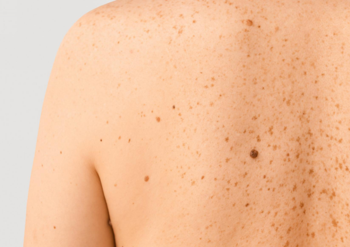
How to Know if a Skin Spot Could Be Skin Cancer
Monitoring changes in your skin is a vital part of maintaining your health. Early identification of skin cancer signs and symptoms allows you to seek timely advice and take positive steps toward ensuring your peace of mind.
Why Do Skin Spots Appear?
Skin spots develop for various reasons, ranging from natural aging to environmental factors. As the body matures, sun exposure triggers the production of melanin, leading to freckles, age spots, and moles. While the majority of these spots are completely benign, ultraviolet (UV) radiation can damage skin cell DNA over time, causing abnormal cell growth that may eventually develop into skin cancer.
What Are the Warning Signs of Skin Cancer?
Regularly checking your skin allows you to become familiar with your baseline moles, making it easier to notice any unusual changes. While many skin developments are harmless, specific visual indicators suggest a spot needs evaluation by a qualified medical professional.
The ABCDE Rule Explained
The ABCDE framework is a widely recognised medical tool used to evaluate whether a mole or spot exhibits characteristics associated with melanoma:
- A – Asymmetry: One half of the spot differs in appearance from the other half.
- B – Border: The edges of the spot are irregular, ragged, notched, or blurred rather than smooth and defined.
- C – Colour: The spot displays multiple shades or an uneven distribution of brown, black, red, white, or blue.
- D – Diameter: The spot is larger than 6mm across (approximately the size of a pencil eraser), though some melanomas can be smaller.
- E – Evolving: The spot undergoes noticeable changes in size, shape, colour, or develops new sensations such as itching, tingling, or bleeding.
Other Warning Signs Beyond ABCDE
Many skin cancers present with features outside the typical ABCDE framework. Other indicators that a spot requires professional assessment include:
- A sore or ulcer that remains open for more than a few weeks.
- A new spot or patch that feels rough, scaly, or develops a crust.
- A small lump that looks shiny, pearly, or translucent.
- Persistent itching, tenderness, or altered sensations in a specific area of the skin.
- A dark streak underneath a fingernail or toenail.
Types of Skin Cancer and How They Look
Different forms of skin cancer manifest with distinct visual characteristics and growth patterns.
Basal Cell Carcinoma (BCC)
Basal Cell Carcinoma is the most frequent form of skin cancer. It commonly presents as a pearly, shiny bump, a smooth pink patch, or a sore that repeatedly oozes and crusts over instead of healing completely.
Squamous Cell Carcinoma (SCC)
Squamous Cell Carcinoma typically develops in areas frequently exposed to the sun, such as the face, ears, and hands. It often looks like a firm, red nodule or a flat, rough spot with a scaly or crusted surface.
Melanoma
Melanoma is a serious form of skin cancer that can develop within an existing mole or appear suddenly as a new spot. It frequently displays the ABCDE characteristics, showing irregular borders, varied colouring, or rapid changes over time.
Skin Spots That Are Usually Harmless
Many spots that appear on the skin are non-cancerous and safe. Common benign spots include:
- Seborrheic Keratoses: Warty, raised growths that often look “stuck on” the skin, varying from light tan to black.
- Cherry Angiomas: Small, smooth, bright red bumps caused by a cluster of capillaries.
- Dermatofibromas: Firm, small brown or pink bumps that typically appear on the legs and dimple inward when pinched.
- Benign Moles (Naevi): Symmetrical, uniform spots with distinct edges that remain stable over time.
Am I at Higher Risk for Skin Cancer?
While anyone can develop skin cancer, certain factors increase individual susceptibility. Understanding these risks can help you determine how vigilantly you should monitor your skin:
- A personal or family history of skin cancer or melanoma.
- A large number of moles or atypical moles (dysplastic naevi).
- Fair skin that burns easily, freckles, or turns red in the sun, along with light-coloured hair or eyes.
- A history of severe, blistering sunburns, particularly during childhood or adolescence.
- Frequent or prolonged exposure to UV radiation from the sun or solariums.
How to Do a Skin Self-Exam at Home
Performing a regular self-examination helps you identify new or changing spots early. It is beneficial to conduct this check once a month in a well-lit room with the assistance of mirrors. Use your mobile phone to photograph any spots or moles that you are concerned about. This can be helpful for comparison if they appear to have changed.
- Check Your Face and Neck: Examine your nose, lips, mouth, and ears using mirrors to view clear angles.
- Inspect Your Scalp: Use a comb or hair dryer to part your hair, checking the scalp section by section.
- Examine Your Upper Body: Look closely at your chest, stomach, shoulders, and underarms.
- Review Your Arms and Hands: Inspect the fronts and backs of your arms, your forearms, palms, backs of hands, and between your fingers.
- Scan Your Back and Lower Body: Use a hand mirror to check your neck, upper back, lower back, and buttocks.
- Check Your Legs and Feet: Examine the fronts, backs, and sides of your legs, your ankles, the soles of your feet, and between your toes.
What Happens at a Professional Skin Cancer Check?
Arranging a professional evaluation is ideal if you notice any new spot that appears distinct from surrounding moles (the “ugly duckling” sign), grows rapidly, oozes fluid, or alters continuously. A professional skin check is a straightforward process focused on thorough examination and accurate identification.
Visual Examination
Biopsy (if needed)
If a spot displays suspicious characteristics, a skin biopsy may be recommended. This involves taking a small sample of the tissue under local anaesthetic and sending it to a pathology laboratory for detailed microscopic analysis.
Results and Next Steps
Following the assessment, the results determine the appropriate pathway. The vast majority of evaluated spots turn out to be completely benign. However, if skin cancer is detected, early identification ensures that surgical removal and other treatment options remain highly effective.
Why Choose Plastic Surgery Central for Skin Cancer Treatment in Adelaide?
For patients requiring surgical removal of a confirmed or highly suspicious skin lesion, Plastic Surgery Central offers a dedicated, patient-centred approach in Adelaide
- Experienced Surgical Care: Consultations and procedures are conducted by our surgeons, who provide surgical options for cosmetic and reconstructive needs
- Accredited On-Site Facilities: Surgical procedures under local anaesthetic can be performed at Central Day Surgery, an accredited on-site day hospital designed for comfort with ground-floor access and adjacent parking
- Collaborative Financial Guidance: Central Day Surgery maintains contracts with select private health insurers to help minimise out-of-pocket costs for skin cancer surgery, assisting patients through the financial aspects of care
- Reconstructive Focus: Our Surgeons focus on precise surgical techniques to support functional recovery and natural-looking structural outcomes after a spot is removed
Remaining attentive to skin changes is an important health habit. If you have identified a spot that concerns you, or if you require an assessment for surgical removal, seeking professional medical advice is the safest next step. Contact our team at Plastic Surgery Central to discuss your options.
Frequently Asked Questions
Can a skin spot look normal and still be skin cancer?
Certain early or clear skin cancers can initially resemble completely normal, harmless spots.
How quickly can a cancerous spot change?
Growth rates vary greatly since slow spots take years, while aggressive melanomas alter within weeks.
Is skin cancer only caused by sun exposure?
Genetics, immune system variations, and chemical exposure also play significant roles in skin cancer development.
How often should I get a professional skin check?
Medical professionals generally recommend scheduling a comprehensive professional skin examination once every twelve months.
Can skin cancer be mistaken for a pimple or an age spot?
Early skin cancers frequently present with features identical to standard pimples, freckles, or aging spots.
What is the difference between a mole and a spot that could be melanoma?
Healthy moles stay uniform and stable, whereas melanomas feature asymmetrical shapes and irregular borders.
Our Surgeons




Recent Post

Thigh Lift Surgery After Weight Loss: What You Need to Know
Saline vs Silicone Breast Implants: Understanding the Differences

Dupuytren’s Contracture: Symptoms, Progression and Treatment Options
Breast Reduction vs Breast Lift: What’s the Real Difference?

Understanding Breast Lift Techniques: What Are the Different Options?
Categories
- Blog (31)
- Body Contouring (2)
- Breast Surgery (8)
- Facial Surgery (7)
- Hand Surgery (4)
- Plastic & Cosmetic Surgery (General) (6)
- Skin & Cancer Surgery (3)